Extracapsular Lateral Suture Stabilization for CCL Injury
Cranial cruciate ligament (CCL) tear is the most common traumatic orthopedic injury seen in dogs of all ages and breeds. The extracapsular lateral suture technique is a stabilization technique that has proven successful in select canine patients, mainly small-to-medium size breeds.
Anatomy of the stifle (knee)
The cruciate ligaments are important stabilizing elements within the canine stifle joint. There are two cruciate ligaments in the knee, called the cranial (anterior) and caudal (posterior) cruciate ligaments. The cranial cruciate ligament (CCL) is commonly injured in both canines and humans (referred to as the ACL in humans).
Effects of CCL rupture
Early signs of CCL stress or partial rupture include stiffness or mild lameness. As the CCL continues to tear further, symptoms increase. A full tear usually results in marked lameness in the affected leg. In some cases, the knee will make a clicking or popping sound as the dog walks. This often indicates damage to the cartilage cushions (menisci) within the knee. When the CCL is ruptured, stifle instability ensues. This instability is often described as cranial tibial thrust or “drawer” movement. This shearing motion causes excessive wear of the cartilage on the ends of the bones within the joint, and stretches the surrounding tissues, causing pain. It can also injure the medial meniscus within the stifle. The goal of stabilization techniques is to eliminate excessive tibial thrust, thus creating a more functionally stable joint and sound gait.
CCL diagnosis
Diagnosis of a CCL tear is made by palpation (feeling the knee) and radiographs (x-rays). The radiographic findings associated with a ruptured CCL include osteoarthritic changes and joint effusion (swelling). The actual ligament cannot be seen on the radiographs.
Description of procedure:
Cranial cruciate ligament (CCL) tear is the most common traumatic orthopedic injury seen in dogs of all ages and breeds. Since the late 1990’s Tibial Plateau Leveling Osteotomy – TPLO, has become a commonly performed technique to address the condition due to its significant success in large and active canine patients.
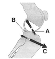
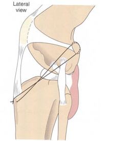
A non-absorbable strong suture material is placed around the lateral fabella and through a small hole created behind the tibial tubercle. The orientation of suture placement attempts to imitate the orientation of the CCL, thus stabilizing the joint. The suture is tightened and tied securely. Two suture strands are typically placed, for added security.
Prognosis with Extracapsular lateral suture stabilization
Most small and medium size dogs return to walking and running after full healing from the procedure. Development of osteoarthritis is still present, and may need to be addressed with conservative measures such as weight control and anti-inflammatory drugs.
Recovery:
Most small and medium size dogs return to walking and running after full healing from the procedure. Development of osteoarthritis is still present, and may need to be addressed with conservative measures such as weight control and anti-inflammatory drugs.