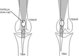
Signs associated with patellar luxation
With lower grades of patellar luxation, dogs display an intermittent lameness and are seen as occasionally “skipping” on the affected limb. As the condition progresses, the dysfunction in the limb becomes more consistent and pronounced, to a point of minimal use and constant abnormal carriage and position of the limb.
Aspects of corrective surgery
Surgical repair may consist of several steps: the kneecap is put back in to the groove, the groove is deepened and the surrounding soft tissues and bone are altered to secure the patella within the groove. Sometime the tibial tuberosity (front section of the shin bone) is transposed to a slightly different location and secured with small pins. If surgery is successful, most dogs return to good use of the limb. Less commonly, in cases where a deformity of the femur (thigh bone) causes the luxation, corrective osteotomy (re-alignment surgery) of this bone is needed.
Surgical aftercare
Strict rest is required following surgery to prevent the patella from re-luxating during the healing process. Initially, there should be no running, jumping, or playing. After a gradual increase in activity, full function is allowed by 2 months in most cases.
Surgical risks or complications
Although uncommon, re-luxation may occur, and often requires an additional surgery. This is of most concern during the first 2 months following surgery, especially in cases with a high grade luxation. If pins were used to transpose the tibial crest, these can sometimes cause irritation and may need to be removed in 3-12 months.
