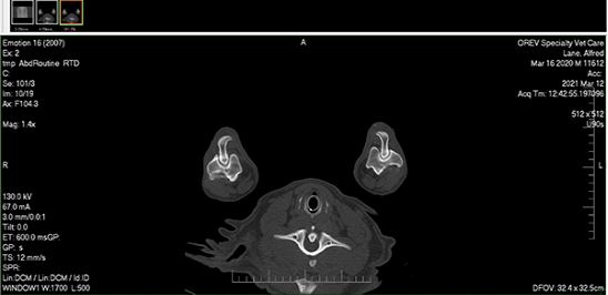
Alfred, a young giant breed dog, came to OREV after having a persistent intermittent forelimb lameness. Exam findings revealed elbow sensitivity, and radiographs showed subtle signs of changes in the elbows. We suspected elbow dysplasia, a hereditary condition causing abnormal growth that affects the elbows. A CT scan of the elbows was recommended to determine whether elbow dysplasia was present, assess the extent of the condition, and tentatively plan surgical treatment.
The CT scan we performed revealed elbow dysplasia in both elbows, with medial coronoid disease (a common component of elbow dysplasia) present. Furthermore, it showed that the right elbow had a fragmented coronoid, and the left elbow had a fissuring of the coronoid. The information was valuable in understanding Alfred's condition, and it allowed us to hone the minimally-invasive arthroscopic surgery that we implemented for Alfred.